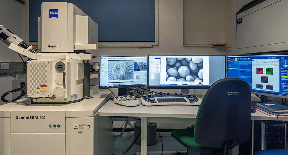
Zeiss GeminiSEM500 Scanning Electron Microscope
The Zeiss GeminiSEM 500 has been installed February 2020 as a versatile, high-resolution system. The microscope has been purchased from a Wellcome Trust grant with Capital Equipment matched funding from the University.
This state-of-the-art scanning electron microscope enables imaging at sub-nanometer resolution with a high detection efficiency especially at low voltages for any type of sample. Crisp images can be acquired fast and with minimum sample damage due to powerful detectors. The new variable pressure mode offers high resolution even with non-coated samples. The Ultim Max100 EDS detector allows for simultaneous electron and x-ray imaging for elemental analysis of samples. With the powerful Zeiss Atlas software large areas can be imaged automatically and correlated with images in multiple dimensions from multiple sources. The system is optimised for array tomography, a volumetric EM method to image large three-dimensional volumes by semi-automated scanning of serial sections.
- Resolution: Up to 0.8 nm at 1 kV
- Accelerating voltage range: 0.02 – 30 kV
- Magnification range: x50 – x2.000.000
- Detection system equipped for parallel detection and signal processing of multiple detector channels
- Chamber detectors and imaging modes:
-SE2 secondary electron detector for standard imaging of surface topography
-VPSE variable pressure SE detector for imaging in variable pressure mode, enables imaging of non-conductive (non-coated) specimen without charging artifacts
-STEM scanning transmission electron microscopy detector
-InLens SE scintillator secondary electron detector for small working distance to image surface structure at high resolution
-Ultim Max 100 Silicon Drift Detector (SDD) EDS detector for elemental analysis with AZtec energy auto system for real-time
chemical imaging
-Chamber CCD camera to monitor position of specimen stage and the distance between objective lens and specimen holder
- Specimen chamber dimensions: 330 mm inner diameter, 270 mm height
- Specimen stage: 5-axes motorised eucentric, controlled from the SmartSEM user interface, operated by dual joystick control box
- Specimen current monitor with integrated touch alarm
- User interface and operating software:
-SmartSEM for general microscope operation
-AZtecLive to operate Ultim Max 100 EDS detector. Combines live electron with live X-ray chemical imaging.
-Zeiss Atlas 5 for multi-scale, multi-modal imaging with a sample-centric correlative environment. Enables automated large area imaging, high throughput and navigation/correlation of images from any source, e.g. light- and x-ray images. Semi-automated imaging of large number of serial sections for array tomography.