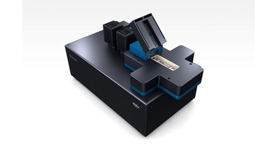
Lightsheet - Luxendo TruLive 3D with Integrated 2Photon Laser-Ablation System
Overview
The Luxendo TruLive 3D Light-Sheet Microscope is a state-of-the-art system equipped with water-based optics, enabling high-resolution imaging of large samples with an extensive usable z-range. This system excels in light-sheet microscopy by employing horizontal illumination, which facilitates rapid z-scanning and significantly reduces photobleaching. This feature is particularly beneficial for prolonged time-lapse imaging, allowing for detailed observation of dynamic biological processes.
A standout feature of our TruLive system is the integration of a high-power 2Photon Coherent laser. This advanced component is adept at activating photo-activatable fluorophores, making it invaluable in cutting-edge fluorescence studies. Moreover, it offers the precision required for single-cell level fluorescence ablation, allowing for meticulous manipulation and study of cellular dynamics.
One of the key innovations of the TruLive system is its rapid-switching mechanism. This feature enables swift and seamless transitions between the 2Photon ablation laser and the light-sheet's imaging lasers. This functionality ensures minimal time delay during operations, enhancing the efficiency of imaging and ablation processes and making the system ideal for intricate and time-sensitive biological research.
- Functional Brain Imaging: Analyzing brain activity and connectivity.
- Large Tissue Imaging: Capturing large tissue volumes with minimal damage.
- Long-term timelapse imaging of a range of model organisms: Spheroids, Blastoids, Zebrafish, and Plants, among others
- Plant Biology: Visualizing internal structures and growth in plants.
- Drug Discovery: Screening drug effects on live cells or organisms.
- Cellular Dynamics: Monitoring intracellular processes and cell division.
- Reduced Photobleaching/Phototoxicity: Minimizes light exposure, preserving sample integrity.
- High Image Quality with Less Haze: Sharp images due to selective thin-section illumination.
- Precise Cell Ablation with 2Photon Laser: Targeted manipulation deep within tissues.
- Rapid Image Acquisition: Fast capture of dynamic biological processes.
- Large Field of View, High Resolution: Comprehensive imaging of large areas in detail.
- Deep Tissue Imaging: Effective for studying thick tissues or whole organisms.
- Compatible with Various Fluorescent Probes: Versatile for multi-color imaging.
- Simplified Sample Preparation: Less complex preparation for thick samples.
- Long-term Live Sample Imaging: Ideal for observing developmental processes over time.
Illumination:
- Dual sided scanned gaussian beam light-sheet illumination,
- Motorised and software-controlled beam geometry (waist, length, position)
- Availability of 6 discrete lasers (405 nm to 685 nm)
- Motorised and software-controlled ND filter set to enable high range dynamic illumination laser power
- MEMS controlled destriping/pivoting
- Motorised cover glass/refractive Index correction
Detection:
- Simultaneous dual spectral channels acquisition for correlative fluorophore analysis, split by dichroic mirrors (3 motorised dichroic mirror positions)
- spectral channel camera alignment
- 20 filter positions split over 2 emission filter wheels, with software-integrated, fast switching between filters
- Motorised and software-controlled magnification changer (positions; 12.5, 25, 37.5 and 50x)
Detection optics include one 25x NA 1.1 water immersion objective lens. The two detection paths are each equipped with ten position high-speed filter wheels, allowing users to collect multiple spectral channels sequentially or dual channel acquisition simultaneously using the motorized dichroic switch carrying three positions. Filter selection includes both long pass-, to maximize photon efficiently, and band pass-filters to minimize bleed through during multi-channel experiments. The detection paths also include a four-position magnification changer that can adjust the overall magnification of the 25x NA 1.1 objective from 12.5x to 62.5x with a click of a button. State of the art Hamamatsu sCMOS Orca Flash 4.0 V3 cameras provide high detection efficiency, quantification, and speed.
Speed and sensitivity range of the camera:
- Maximum frame rate >80 fps at full frame (2048 × 2048 pixels of 6.5 μm × 6.5 μm size) and up to 500 fps at subframe cropping
- Peak quantum efficiency (QE): 82% @ 560 nm
- Two spectral detection channels, each equipped with a fast filter wheel (10 positions and 50 ms switching time between adjacent positions) Filters adapted to the selected laser lines
Laser Lines:
- 405nm Blue DAPI / Alexa 350
- 488nm Green GFP / Alexa 488 / FITC
- 561nm Red DsRed / TagRFR / Alexa 568
- 642nm Far Red Alexa 633 / CY5
- 685nm Far Red Alexa 633 / CY5
Incubation:
- Incubation with gas control (CO2, O2, humidity), with humidity sensor in closest proximity to the sample for accurate gas mixture modulation
- Incubation with temperature control (25-45 °C), with sensors in closest proximity to the sample for fast temperature modulation (5°C/min) and closed feedback cycle
Photomanipulation module:
Visible CW lasers (405 nm – 780 nm) for photoconversion, FRAP and optogenetics, etc.
Pulsed lasers: Coherent Chameleon Vision II 680-180nm; tunning speed >40 nm s-1 ; Pulse width 140±20 fs.
TruLive3D Dishes:
The TruLive3D Dishes are ready-to-use sample holders for the Luxendo TruLive3D Imager light-sheet microscopes. The TruLive3D Dishes have been sterilized and are shipped in sealed sleeves. To retain sterile conditions, open the packaging sleeve only under a cell culture hood. Similar to the classic sample holder, the FEP foil lining the holder serves the dual function of a “curved coverglass” and a physical barrier between the immersion medium and sample medium. The new ready-to-use TruLive3D Dishes are fully compatible with the TruLive3D Imager. TruLive Dishes enable the parallel imaging of samples grown under different media conditions.


