Facilities
Facility equipment
Take a look at the information below to find out more about the equipment that we are able to offer as well as our sample preparation facilities:
Narrowband Spectrally Focused Coherent Raman
This custom-built system provides fast hyperspectral CRS imaging using chirped femtosecond laser pulses to achieve rapid Raman spectra acquisition while retaining the full speed and image quality of narrowband SRS imaging.
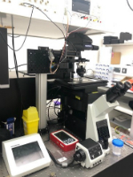
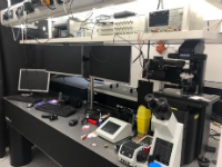
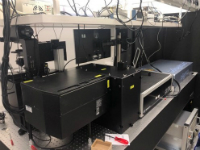
This system is powered by an InSightX3 fs laser, which provides a tuneable output between 680-1300nm and a fixed wavelength output at 1045nm. The 2 beams are combined in the SF-TRU spectral focusing unit. This unit controls the laser powers applied to the sample, the time delay between the beams. It also provides the intensity modulations in the Stokes beam necessary for SRS and chirps the beams from fs to ps pulses. The imaging is carried out on a modified confocal inverted microscope (Olympus Flouview3000 and IX81). The light is focused on the sample with a 1.2NA water objective and transmitted light is collected with either an oil or water condenser depending on the sample type. For transparent samples SRS and CARS are measured in the forwards direction, however for thicker samples there is also an option to collect backscattered CARS in an epi channel. In this lab we have the ability to combine Coherent Raman imaging with complementary techniques including Second Harmonic Generation SHG and Two Photon fluorescence (TPF), which will provide complementary information about the collagen structures and endogenous fluorophores or added fluorescent labels within a sample. The system is set up for 3D imaging, large area scans and rapid hyperspectral imaging.
Further reading:
- Module for multiphoton high-resolution hyperspectral imaging and spectroscopy (Aram Zeytunyan; Tommaso Baldacchini; Ruben Zadoyan) - February 2018
 |
 |
 |
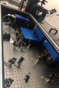
Broadband CARS Microscope
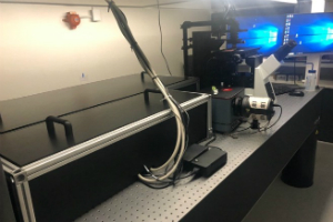
This custom-built microspectrometer provides two- and three-dimensional mapping of biomolecules based on their Raman fingerprint. Based on broadband coherent anti-Stokes Raman scattering this system provides a combination of speed, sensitivity and spectral breadth. The system utilizes a configuration of laser sources that probes the entire biologically relevant Raman window (500–3500 cm–1). By utilising intrapulse three-colour excitation and the non-resonant background to heterodyne-amplify weak Raman signals it is possible to obtain efficient acquisition of the typically weak ‘fingerprint’ region.
At the heart of the system are two co-seeded fibre lasers (Toptica, FemtoPro), custom designed specifically for broadband CARS. The system provides synchronization of narrowband (∼3.4 ps) flat-top pulses at 770 nm synchronised with supercontinuum pulses (∼16 fs) spanning ∼900–1400 nm. The first fibre laser, a FemtoFiber pro NIR is a frequency doubled ultrafast Erbium fibre laser coupled to a FF 1PS NIR FemtoFiber pro to increase the pulse length to ~3.4 ps. The second fibre laser is an ultrashort-pulsed Erbium fibre amplifier (FF PRO UCP AMP) with motorised control for optimisation of the supercontinuum power and pulse duration. The FF PRO UCP AMP is seeded by the FF PRO NIR so that the two pulse trains are intrinsically rep-rate locked.
The beams are temporally and spatially overlapped and delivered onto the sample via a 1.2NA objective and the CARS signal collected in the forwards direction by and inverted microscope (Olympus IX73) with a 0.7NA objective. The anti-Stokes signal is spectrally isolated from the excitation wavelengths using band-pass filters and focused onto the entrance slits of a spectrograph (Princeton Instruments Isoplane 160) equipped with a fast CCD camera (Princeton Instruments Pixis).
Further reading:
- High-speed coherent Raman fingerprint imaging of biological tissues (Charles H. Camp Jr, Young Jong Lee, John M. Heddleston, Christopher M. Hartshorn, Angela R. Hight Walker, Jeremy N. Rich, Justin D. Lathia & Marcus T. Cicerone) - July 2014
 |
 |
Support for Data Analysis
At the CONTRAST facility we also offer support for analysing the large volumes of data generated by the system.
For example Hyper-spectral image data can be analysed by a multivariate curve resolution methods to reconstruct quantitative concentration images for each individual component and retrieve the corresponding vibrational Raman spectra.
For 3D and large area maps we can also reconstruct images which can be compared to traditional H&E staining by obtaining images taken at a few key Raman shifts.
Sample Preparation facilities
CONTRAST facility users will have access to a shared cell culture laboratory. We have a class II biosafety facilities however do not routinely handle pathogenic, infectious or genetically modified organisms therefore work involving such agents would require discussion with the team.
We can image both fixed and living cells on the coherent Raman microscopes. Living cells are best imaged in 1inch diameter well plates with a coverslip bottom.
We are able to work with Human tissue samples at the facility. However before receiving these and allocating microscopy time we will need to have copies of all the ethical paper work including ethical approval for the samples to be transported to our site. Additionally we will require a material transfer agreement for the samples. Human tissue samples can be stored in our -80 freezer prior to investigation, however we will need to keep accurate records of all HTA licensable and non-licensable samples and we limit the time samples can be stored to a maximum of 3 months.
Imaging tissue sections: Tissue sections can be any thickness as long as they are still translucent and light can reach the detectors in the forwards direction. Samples are best mounted between 2 glass coverslips as this allows the optimal amount of light to reach the detectors, however we can also handle samples mounted between a microscopy slide and a cover slip.
Cell Imaging: We can image both fixed and living cells on this system. For living cells researchers also have access to our cell culture laboratory. Living cells are best imaged in at least 1inch diameter well plates with a coverslip bottom.
Samples can be prepared either fresh or fixed and there is no need to add exogenous labels, however on the spectrally focused SRS system, we can combine coherent Raman imaging with TPF imaging of fluorescent labels if this is required.
Other samples: If you have a different kind of sample you would like to image please contact us, we will be able to do a trail imaging session to determine the best sample preparation technique.
